Embolic Microspheres are compressible hydrogel microspheres with a regular shape, smooth surface, and calibrated size, which are formed as a result of chemical modification on polyvinyl alcohol (PVA) materials. Embolic Microspheres consist of a macromer derived from polyvinyl alcohol (PVA), and are hydrophilic, non-resorbable, and are available in a range of sizes. The preservation solution is 0.9% sodium chloride solution. The water content of fully polymerized microsphere is 91% ~ 94%. Microspheres can tolerate compression of 30%.
Embolic Microspheres are intended to be used for the embolization of arteriovenous malformations (AVMs) and hypervascular tumors, including uterine fibroids. By blocking the blood supply to the target area, the tumor or malformation is starved of nutrients and shrinks in size.
In this article, we will show you the detailed steps about how to use Embolic Microspheres.
Goods preparation
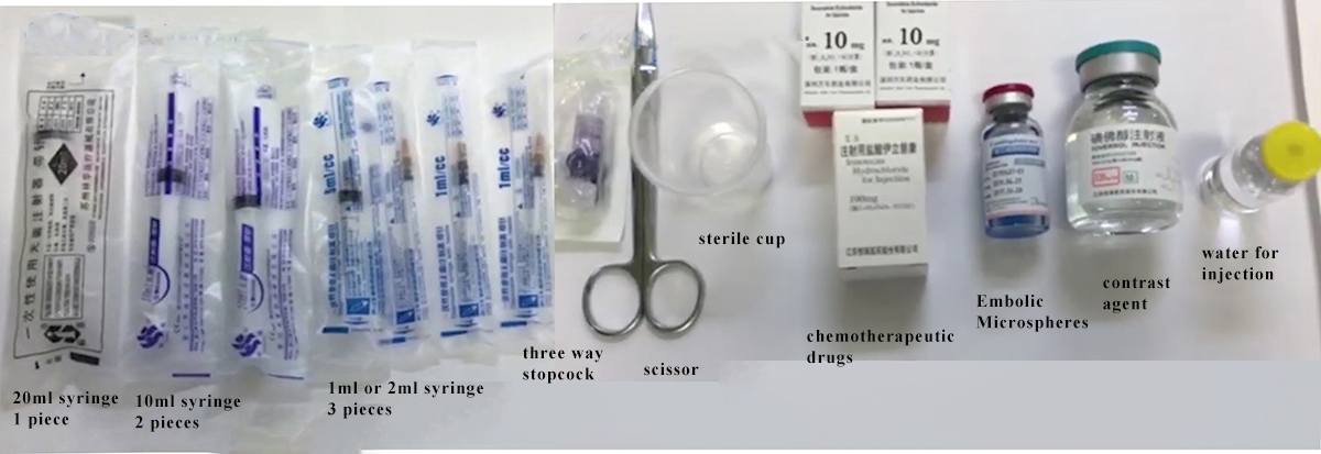
It is necessary to prepare 1 20ml syringe, 2 10ml syringes, 3 1ml or 2ml syringes, three-way, surgical scissors, sterile cup, chemotherapy drugs, embolic microspheres, contrast media, and water for injection.
Step 1: Configure chemotherapy drugs
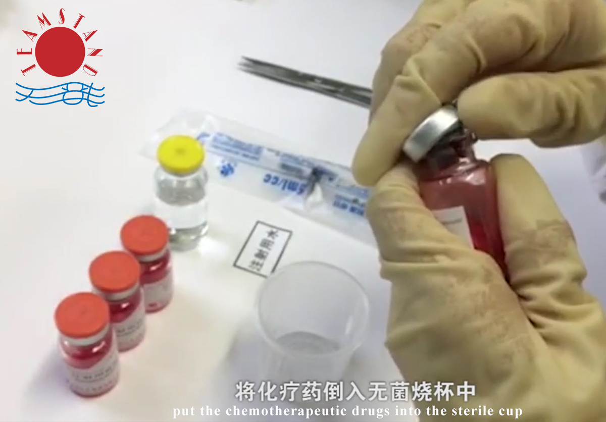
Use surgical scissors to uncork the chemotherapeutic medicine bottle and pour the chemotherapeutic medicine into a sterile cup.
The type and dosage of chemotherapeutic drugs depend on clinical needs.
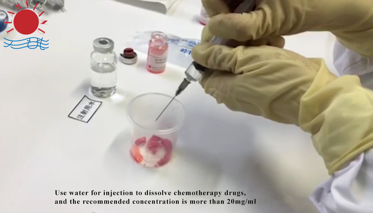
Use water for injection to dissolve chemotherapy drugs, and the recommended concentration is more than 20mg/ml.
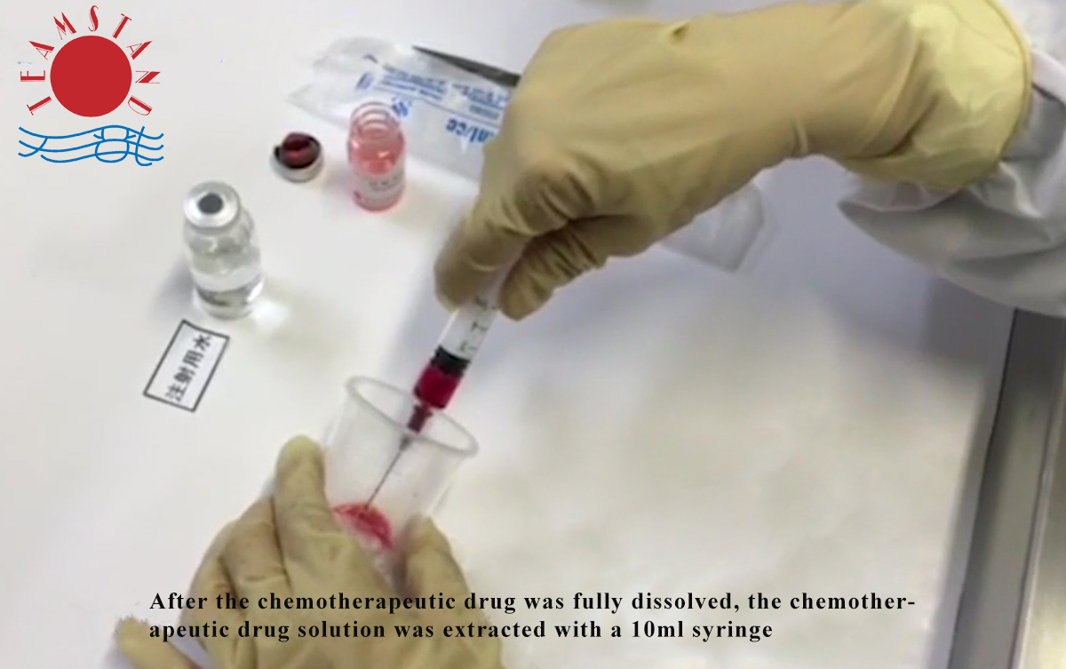
After the chemotherapeutic drug was fully dissolved, the chemotherapeutic drug solution was extracted with a 10ml syringe.
Step 2: Extraction of drug-carrying embolic microspheres
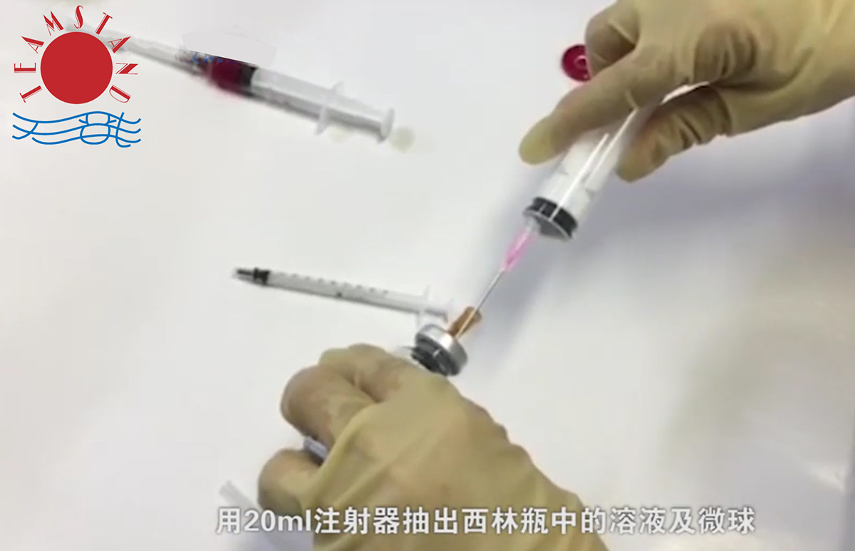
The embolized microspheres were fully shaken, inserted into a syringe needle to balance the pressure in the bottle, and extract the solution and microspheres from the cillin bottle with a 20ml syringe.
Let the syringe stand for 2-3 minutes, and after the microspheres settle, the supernatant is pushed out of the solution.
Step 3: Load the Chemotherapeutic drugs into Embolic Microspheres
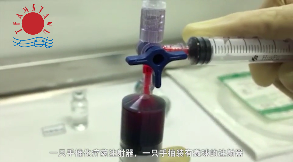
Use the 3 ways stopcock to connect the syringe with the embolic microsphere and the syringe with the chemotherapy drug, pay attention to the connection firmly and the flow direction.
Push the chemotherapy drug syringe with one hand, and pull the syringe containing embolic microspheres with the other hand. Finally, the chemotherapy drug and microsphere are mixed in a 20ml syringe, shake the syringe well, and leave it for 30 minutes, shake it every 5 minutes during the period.
Step 4: Add contrast media
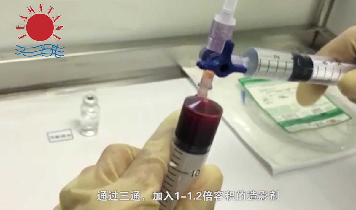
After the microspheres were loaded with chemotherapeutic drugs for 30 minutes, the volume of the solution was calculated.
Add 1-1.2 times the volume of contrast agent through the three way stopcock, shake well and let stand for 5 minutes.
Step 5: Microspheres are used in the TACE process
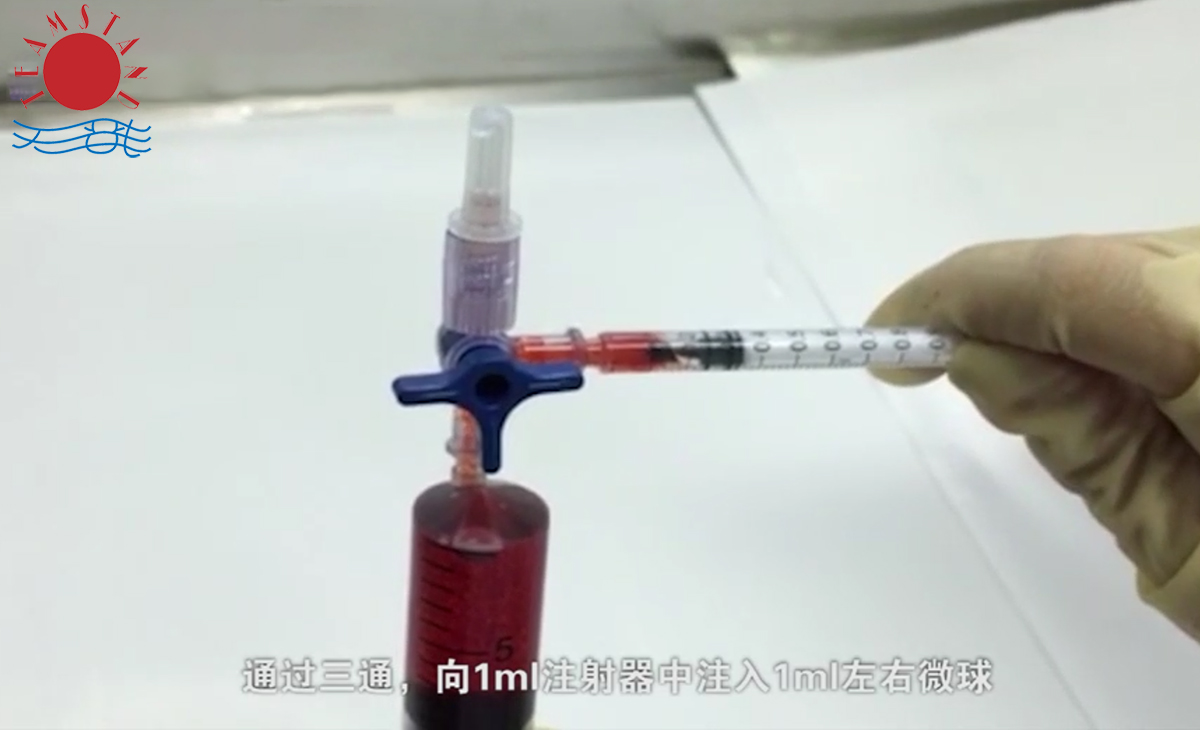
Through the three way stopcock, inject about 1ml of microspheres into the 1ml syringe.
The microspheres were injected into the microcatheter by pulsed injection.
Guides attentions:
Please ensure aseptic operation.
Confirm that the chemotherapeutic drugs are completely dissolved before loading the drugs.
The concentration of chemotherapy drugs will affect the drug loading effect, the higher the concentration, the faster the adsorption rate, the recommended drug loading concentration is not less than 20mg/ml.
Only sterile water for injection or 5% glucose injection should be used to dissolve chemotherapy drugs.
The rate of doxorubicin dissolution in sterile water for injection was slightly faster than 5% glucose injection.
5% glucose injection dissolves pirarubicin slightly faster than sterile water for injection.
The use of ioformol 350 as contrast medium was more conducive to the suspension of microspheres.
When injected into the tumor through microcatheter, pulse injection is used, which is more conducive to microsphere suspension.
Post time: Feb-28-2024


